
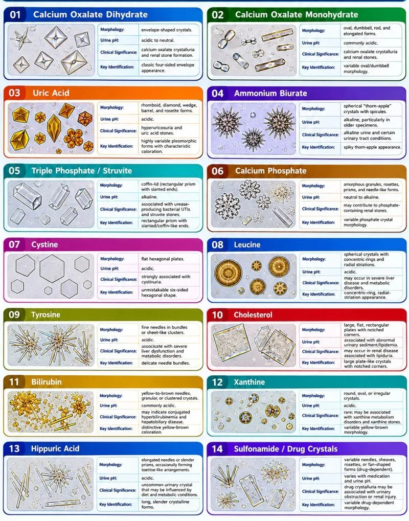
Introduction
Urinary crystals are microscopic structures formed when substances dissolved in urine become sufficiently concentrated or supersaturated and precipitate into solid particles.
They are commonly encountered during microscopic examination of urinary sediment and can provide useful information about the chemical environment of urine, metabolic conditions, medications, urinary tract infections, and potential stone formation.
However, an important principle in urinalysis is that the presence of urinary crystals does not automatically indicate disease.
Some crystals may occur in healthy individuals, particularly when urine is concentrated or when specimen conditions favor precipitation. Other crystal types are more clinically significant and may point toward specific metabolic or pathological conditions.
How Do Urinary Crystals Form?
Crystal formation occurs when urine becomes supersaturated with substances capable of precipitating.
Several factors influence this process.
1. Urine Concentration
Concentrated urine contains higher levels of dissolved substances. Dehydration can therefore increase the concentration of substances such as calcium, oxalate, uric acid, and phosphate and may favor crystallization.
2. Urine pH
Urine pH strongly influences which substances remain soluble.
Some crystals are more likely in acidic urine, while others are favored by alkaline conditions.
For example:
- Uric acid crystals → commonly associated with acidic urine.
- Struvite crystals → commonly associated with alkaline urine.
3. Temperature
Temperature can influence solubility. Cooling a urine specimen may cause certain substances to precipitate, potentially producing crystals that were not prominent in the freshly voided sample.
4. Specimen Storage and Delay
Urine sediment can change over time. Delayed examination may alter pH, bacterial growth, cellular preservation, and crystal formation.
For this reason, proper specimen handling and timely examination are important when interpreting urinary crystals.
5. Diet and Metabolism
Dietary intake and metabolic disorders can influence urinary concentrations of substances involved in crystallization.
Examples include changes in uric acid, calcium, oxalate, phosphate, and cystine excretion.
6. Medications
Some medications and their metabolites may precipitate in urine and appear as crystals. When unusual crystals are identified, medication history can therefore provide an important clue.
7. Infection
Certain urinary tract infections can alter urine chemistry and promote crystal formation.
Urease-producing bacteria can increase urinary ammonia and raise urine pH, creating conditions that favor magnesium ammonium phosphate (struvite) crystallization.
Common Urinary Crystals
Calcium Oxalate
Calcium oxalate is one of the most frequently encountered urinary crystals.
The classic form has an envelope-like appearance, although other forms such as dumbbell or ovoid shapes may occur depending on the crystal type and conditions.
Calcium oxalate crystals may occur in normal urine, particularly when the urine is concentrated. They are also associated with calcium oxalate urinary stones.
The presence of calcium oxalate crystals alone, however, does not prove that a patient has kidney stones.
Uric Acid
Uric acid crystals are usually found in acidic urine and may demonstrate several morphological forms, including:
- Rhomboids
- Diamonds
- Rosettes
- Barrels
- Wedges
Persistent uric acid crystallization can be associated with increased uric acid concentration and may contribute to uric acid stone formation.
Struvite
Struvite crystals consist of magnesium ammonium phosphate and are typically found in alkaline urine.
Their classic appearance is described as a coffin-lid shape.
Struvite crystallization is strongly associated with urinary infections involving urease-producing organisms. These organisms can increase urine alkalinity and promote precipitation of magnesium ammonium phosphate.
Cystine
Cystine crystals are characteristically hexagonal.
Their identification is particularly important because cystine crystalluria is strongly associated with cystinuria, an inherited disorder involving impaired renal reabsorption of certain amino acids.
Cystine crystals are therefore an example of a urinary sediment finding that can provide an important diagnostic clue.
Other Crystals of Clinical Interest
Although calcium oxalate, uric acid, struvite, and cystine are commonly emphasized, other crystals may also be encountered.
These include various phosphate crystals, amorphous urates, amorphous phosphates, and crystals associated with medications or metabolic abnormalities.
The appearance of a crystal should therefore always be interpreted alongside urine pH, specimen conditions, and clinical information rather than relying on morphology alone.
Crystalluria Does Not Equal Urolithiasis
One of the most important distinctions in urinary microscopy is the difference between crystalluria and urolithiasis.
Crystalluria means crystals are present in the urine. Urolithiasis refers to the formation of a clinically relevant urinary stone.
A patient can have crystalluria without having a urinary stone, and the presence of crystals alone cannot establish a diagnosis of nephrolithiasis.
However, persistent or abundant crystalluria may indicate a urinary environment that favors stone formation and may be relevant when interpreted together with symptoms, biochemical findings, and imaging.
Laboratory Identification
When examining urinary sediment, the medical laboratory scientist should consider several factors rather than relying solely on the shape of a crystal.
Important considerations include:
- Crystal morphology
- Urine pH
- Specific gravity or concentration
- Specimen age
- Storage temperature
- Associated cells and casts
- Chemical urinalysis findings
- Patient history
- Medication history
- Previous urinary stone history
Polarized microscopy can provide additional information for certain crystals, while confirmatory testing may be required when a specific metabolic disorder or unusual crystal is suspected.
Why Specimen Handling Matters
A freshly collected urine specimen can change considerably if it is left standing for an extended period.
Bacterial multiplication may alter urine pH, cells may deteriorate, and changes in temperature can affect the solubility of urinary substances.
Consequently, laboratory scientists should follow the laboratory's validated requirements for specimen storage and examination time.
This is especially important when crystal morphology is being used to support clinical interpretation.
Clinical Interpretation
Not all crystals require clinical intervention. Some may represent incidental findings caused by concentration, diet, specimen conditions, or transient changes in urine chemistry.
Further investigation may be appropriate when crystalluria is:
- Persistent
- Abundant
- Associated with hematuria
- Associated with recurrent urinary stones
- Accompanied by renal dysfunction
- Associated with urinary obstruction
- Associated with significant urinary symptoms
- Suggestive of a metabolic disorder
- Suggestive of drug-induced crystalluria
The laboratory finding should therefore be interpreted as part of the overall clinical picture.
Quick Identification Guide
| Crystal | Typical Urine Environment | Characteristic Appearance | Important Association |
|---|---|---|---|
| Calcium Oxalate | Variable | Envelope/dumbbell forms | Calcium oxalate stones |
| Uric Acid | Acidic | Rhomboid/diamond/rosette forms | Uric acid stone formation |
| Struvite | Alkaline | Coffin-lid | Urease-producing urinary infection |
| Cystine | Usually acidic | Hexagonal | Cystinuria |
Key Takeaway
Urinary crystals are clues, not diagnoses. Their morphology, urine chemistry, quantity, specimen conditions, and clinical context must all be considered before determining their significance.
For the medical laboratory scientist, good urinary microscopy is therefore more than recognizing shapes. It involves understanding why the crystal formed, whether the specimen conditions could have contributed to its formation, and what the finding may mean in the clinical context.