
Image Attributions
Images used in LabGOTO assessment questions are sourced from public-domain and Creative-Commons licensed repositories. Credits are listed below.
Wikimedia Commons

Gram-positive cocci arranged in clusters are most consistent with which organism?
Wikimedia Commons · CC BY-SA 4.0 · Source

Which rapid test detects acid-fast bacilli and is commonly used for TB?
Wikimedia Commons · CC BY-SA 4.0 · Source

The peripheral smear demonstrates red cells with a bull's-eye appearance: a central button of hemoglobin, a pale ring, a...
Wikimedia Commons · CC BY 2.0 · Complete on fetch: author via Wikimedia Commons, CC BY-SA 4.0 · Source

The smear demonstrates erythrocytes with a slit- or mouth-shaped area of central pallor. Which morphology is shown?
Wikimedia Commons · CC BY 4.0 · Complete on fetch: author via Wikimedia Commons, CC BY-SA 4.0 · Source

The smear demonstrates crescent-shaped, elongated red cells with pointed ends. These cells are most associated with whic...
Wikimedia Commons · CC BY-SA 4.0 · Complete on fetch: author via Wikimedia Commons, CC BY-SA 4.0 · Source
The smear demonstrates fragmented, helmet-shaped red cells. This finding is most consistent with which process?
Wikimedia Commons · CC BY 4.0 · Complete on fetch: author via Wikimedia Commons, CC BY-SA 4.0 · Source
The Gram stain demonstrates Gram-positive cocci arranged in clusters. Which organism is most likely?
Wikimedia Commons · CC BY-SA 4.0 · Courtesy of CDC Public Health Image Library (public domain) · Source

The Gram stain demonstrates lancet-shaped Gram-positive diplococci. Which organism is most likely?
Wikimedia Commons · CC0 · Courtesy of CDC Public Health Image Library (public domain) · Source

A stool specimen demonstrates a pear-shaped flagellate with two nuclei giving a falling-leaf appearance on wet mount. Wh...
Wikimedia Commons · CC BY-SA 4.0 · Courtesy of CDC DPDx (public domain) · Source

A cornmeal agar preparation demonstrates yeast cells with pseudohyphae and blastoconidia. Which organism is most likely?
Wikimedia Commons · CC BY-SA 4.0 · Courtesy of CDC Public Health Image Library (public domain) · Source

The smear shows small, round, dense red cells lacking central pallor. Which morphology is shown?
Wikimedia Commons · CC BY-SA 4.0 · Complete on fetch: author via Wikimedia Commons, CC BY-SA 4.0 · Source

A urethral smear shows Gram-negative diplococci within neutrophils. The most likely organism is:
Wikimedia Commons · CC BY 4.0 · Courtesy of CDC Public Health Image Library (public domain) · Source
A Ziehl-Neelsen stain shows red, slender acid-fast bacilli. The organism is most likely:
Wikimedia Commons · CC BY-SA 4.0 · Courtesy of CDC Public Health Image Library (public domain) · Source

Urine microscopy shows casts containing white cells. These casts indicate:
Wikimedia Commons · CC BY-SA 4.0 · Complete on fetch: author via Wikimedia Commons, CC BY-SA 4.0 · Source
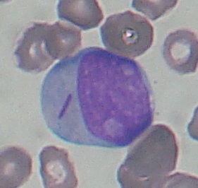
A blast on the smear contains a rod-shaped, azurophilic inclusion. This finding supports:
Wikimedia Commons · CC BY-SA 3.0 · Complete on fetch: author via Wikimedia Commons, CC BY-SA 4.0 · Source

The smear shows a bilobed cell with large orange-red granules. This cell is increased in:
Wikimedia Commons · CC BY-SA 4.0 · Complete on fetch: author via Wikimedia Commons, CC BY-SA 4.0 · Source

Urine microscopy shows colorless prisms with a coffin-lid appearance. These crystals are:
Wikimedia Commons · CC BY-SA 3.0 · Complete on fetch: author via Wikimedia Commons, CC BY-SA 4.0 · Source